How Is AI different from a breast density assessment?
As a patient, you might have received a category of breast density on your screening mammogram report. This probably means nothing to you except that you got a negative screening mammogram, however you may want an explanation. To do so, we need to explain what makes the breast tissue. The breast consists of milk glands, milk ducts and thicker tissue referred to as supportive tissue and fatty tissue – see illustration below. Dense tissue consists of the milk glands, milk ducts and the supportive tissue. It is thicker and more difficult to penetrate via x-rays. Fat is very loose and easily penetrated with x-rays.
When a mammogram is performed the compression paddle tries to flatten out the breast making it even in thickness and to help tissue to spread out so as to not be superimposed allowing the x-rays to penetrate the tissue. Dense tissue may flatten out but remains thicker and fibrous, while showing up white on the mammogram. Fat would show up dark on the mammogram and be very easy to see through.
Breast Density
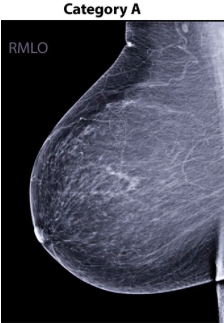
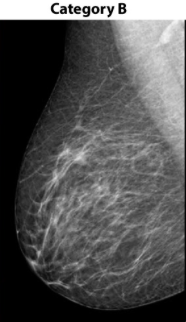
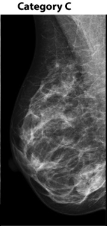
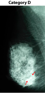
Let’s define breast density. Four density categories (A to D) were developed to provide a consistent interpretation of mammograms and establish a standard of treatment. These categories are as follows(1):
A) Fatty – very easy to see through on x-ray
B) Scattered fibroglandular density – dense areas scattered throughout the breast but mostly fat
C) Heterogeneously dense – a lot of the breast is thick tissue and has less fat which makes it harder to see through with x-ray
D) Extremely dense – most of the breast is thick tissue and contains very little fat, which makes it very difficult to see through with x-ray.
Breast density can be an issue as a negative mammogram could easily have cancerous lesions hidden in a category C or D (see below category D). Breast density has been, and remains, a visual assessment. However, technology could help.
Now, breast density can be categorized with the assistance of software programs that are written specifically to quantify it. The software focuses on the specific types of tissue to give a more objective assessment of the density. This can be integrated with the risks of personal history to assist physicians in a more personalized approach to screening, and possible earlier detection of cancer. These breast density softwares do not assist with the mammographic assessment nor with the detection of suspicious lesions.
How is AI for mammography different?
AI for mammography is designed to assess the entire mammogram, regardless of the density. The analysis comes from the images, with the focus on detection of early cancer. AI for mammography improves workflow, reduces call backs for additional imaging and improves cancer detection. MammoScreen® AI points only to the most suspicious findings and their location in the mammogram. This allows the physician to not only navigate quickly to the most relevant area of interest but also offers full traceability and explanation of the AI’s opinion. By pointing out suspicious lesions, or bringing reassurance in benign cases, the radiologists’ accuracy and workflow is improved.
Overall Summary
Software is used for both breast density and mammogram assessments. The difference lies in how they are used. Breast density is an issue of assessing the type of tissue that the patient has in order to assess the breast cancer risk. Mammogram assessment is a matter of detecting and characterizing the suspicious lesions visible on the images.
Artificial intelligence for mammography is used to relieve the high demand workflow that radiologists incur and to better assess the mammograms for early cancer detection. It provides the radiologist with a second pair of eyes. AI alone is good – AI and radiologists are better together. Studies have shown (2) that the best results are obtained when radiologists utilize the power of AI to help optimize accuracy and performance for breast cancer screening.
Learn more about Artificial Intelligence and Mammography by visiting our patient information page.
(1)https://densebreast-info.org/for-patients/
(2) https://www.itnonline.com/article/exciting-future-ai-and-mammography-and-physicians