Case of the week (week 11, 2022)
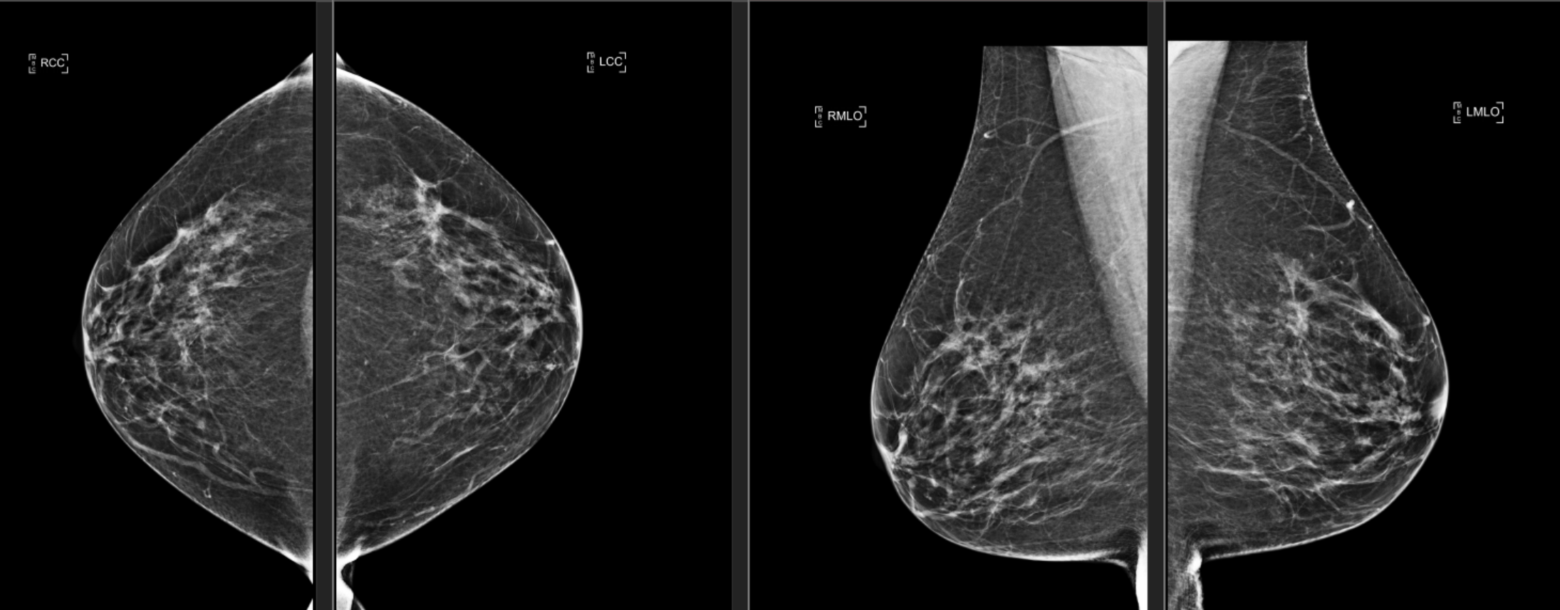
BILATERAL SCREENING 3D MAMMOGRAM
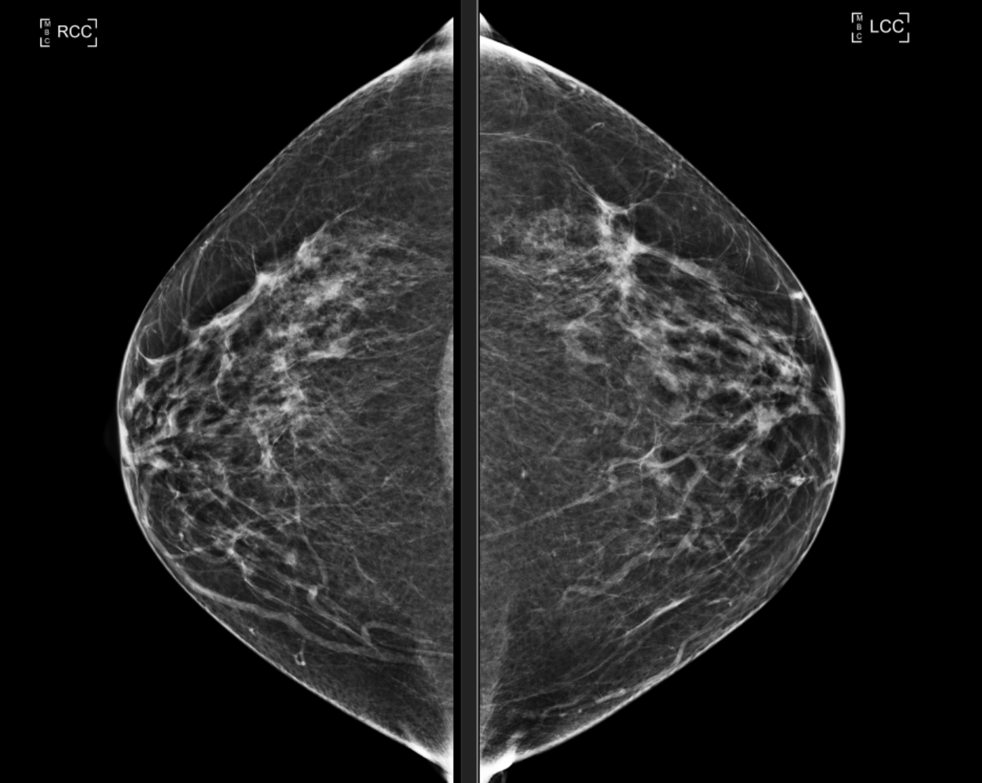
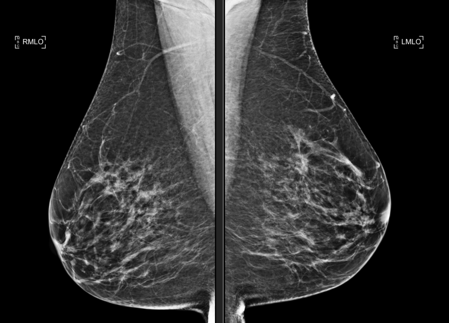
The breasts are heterogeneously dense. In the left upper outer quadrant middle third, there is architectural distortion. No significant change in the appearance of the right breast, with no new dominant mass lesions, suspicious microcalcifications or areas of architectural distortion.
Using MammoScreen®, a co-located mass with microcalcifications in the LEFT CC and LEFT MLO with a MammoScreen ScoreTM of 9 was found. Right breast scored a 1 – no findings.
MAMMOGRAM FINDINGS: There is an irregular spiculated 1.5 cm mass left breast 2:00 position 5 cm from the nipple. This is most apparent on 3D images.
ULTRASOUND FINDINGS: In the left breast 2:00 position 5 cm from the nipple posterior depth there is an irregularly marginated solid densely shadowing 1.7 x 1.5 x 1.6 cm mass. There is some peripheral vascularity. The targeted breast exam tissue at this site is firm. No suspicious left axillary adenopathy.