Case of the week (week 12, 2022)
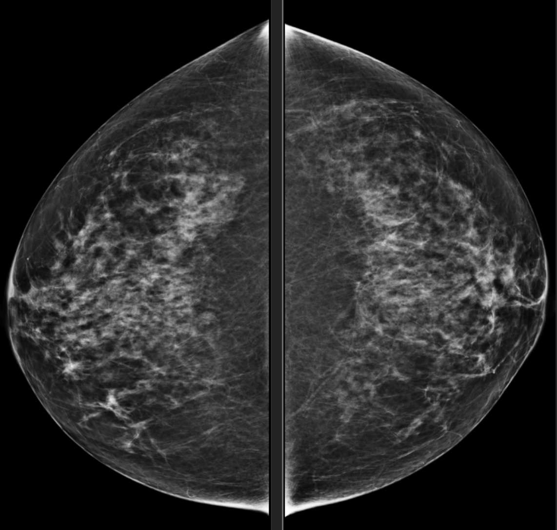
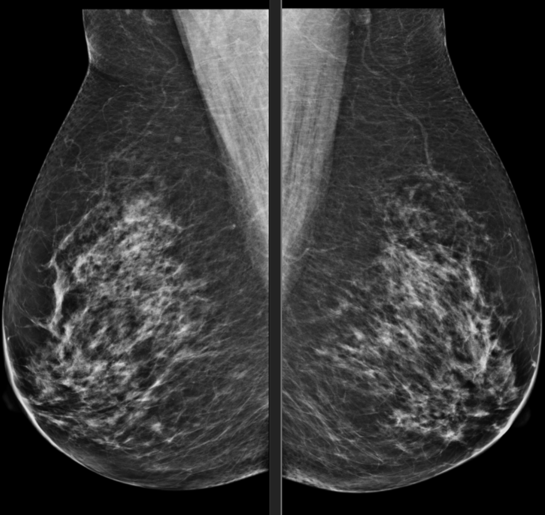
BILATERAL SCREENING 3-D MAMMOGRAM
Heterogeneously dense (50 to 75%) breasts. There is an area of subtle questionable architectural in the upper central left breast posterior depth. Additional imaging is recommended for further evaluation.
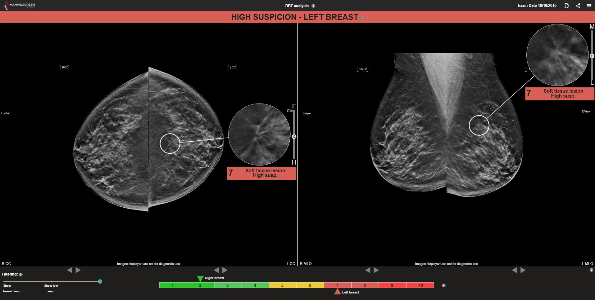
MammoScreen®: A soft tissue lesion in the LEFT CC and LEFT MLO are indicated with a MammoScreen ScoreTM of 7. Right breast scored a 2 – lowest suspicion.
MAMMOGRAM FINDINGS: Breast tissue is heterogeneously dense. A true lateral and 2 focal compression were performed and 3D tomosynthesis. On the additional views, best demonstrated with 3D tomosynthesis images is a subtle area of architectural distortion in the 12:00 position centered about 8 cm from the nipple. No dominant mass or suspicious microcalcification elsewhere in the left breast.
ULTRASOUND FINDINGS: In the 12:00 position centered about 8-9 cm from the nipple is a focal area of irregular shadowing without a well-circumscribed mass. On directed physical exam, breast tissue is slightly nodular but no clearly fixed firm mass on exam.
Biopsy results confirmed low grade invasive ductal carcinoma.