Case of the week (week 3, 2021)
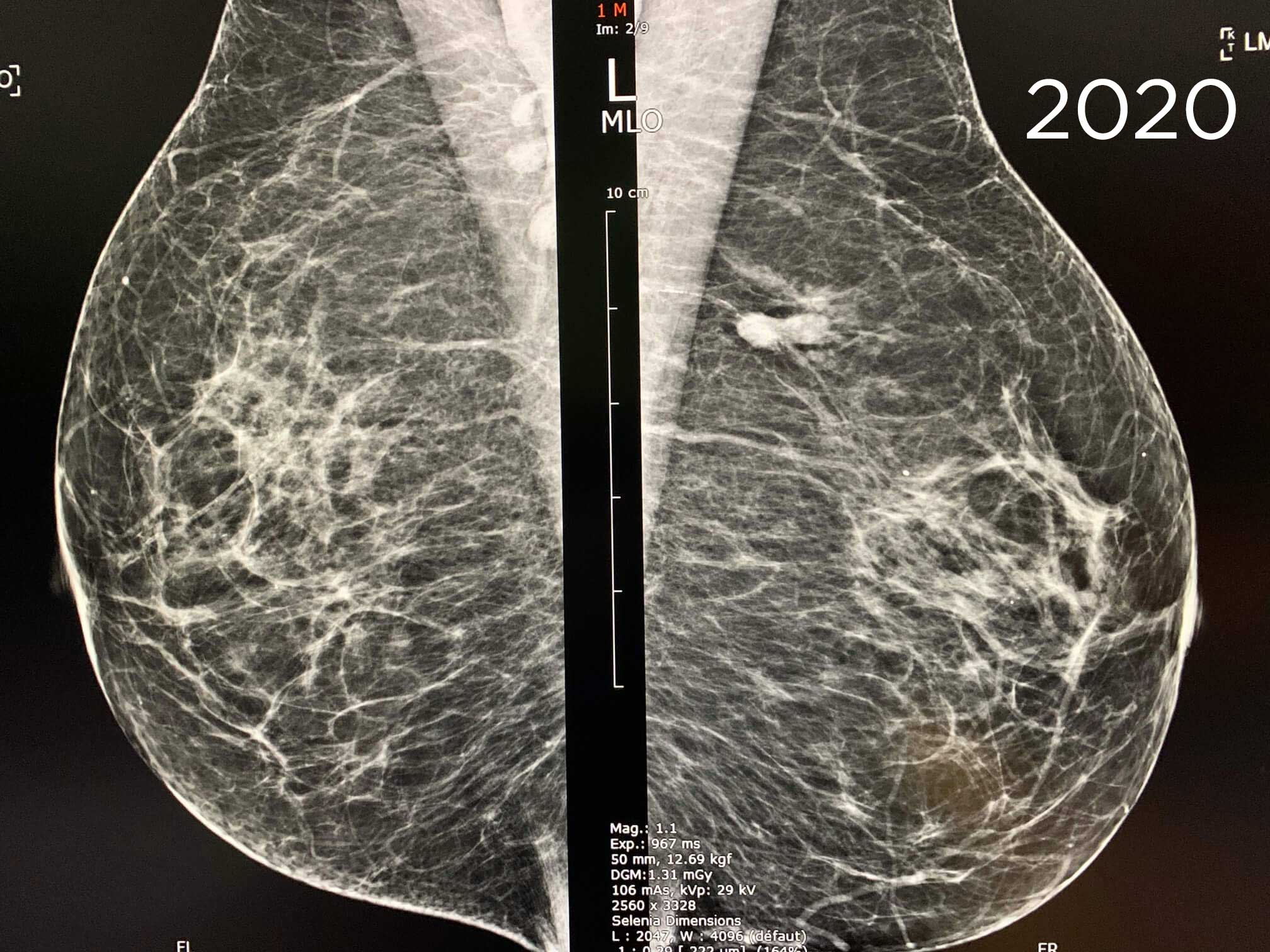
Screening mammography. 73 year-old patient.
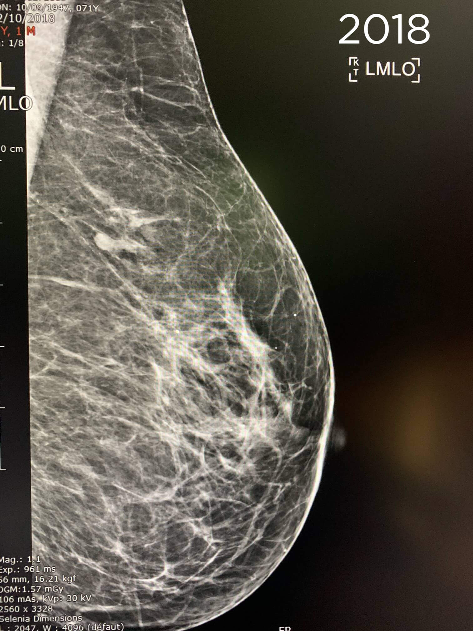
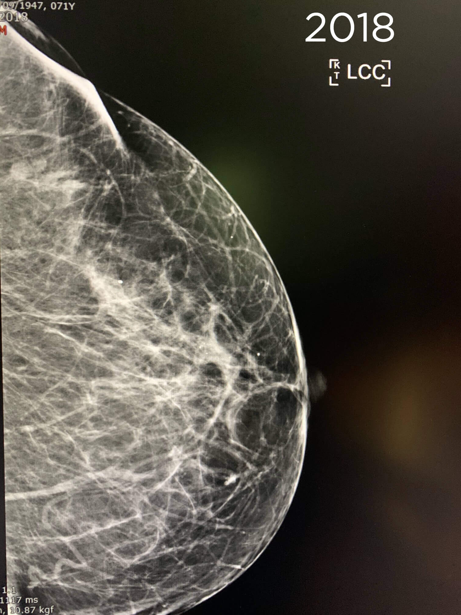
The mammography is compared to the prior of 2018. 2 fused opacities of the left axillary extension were present on the prior of 2018 evoking intramammary lymph nodes.
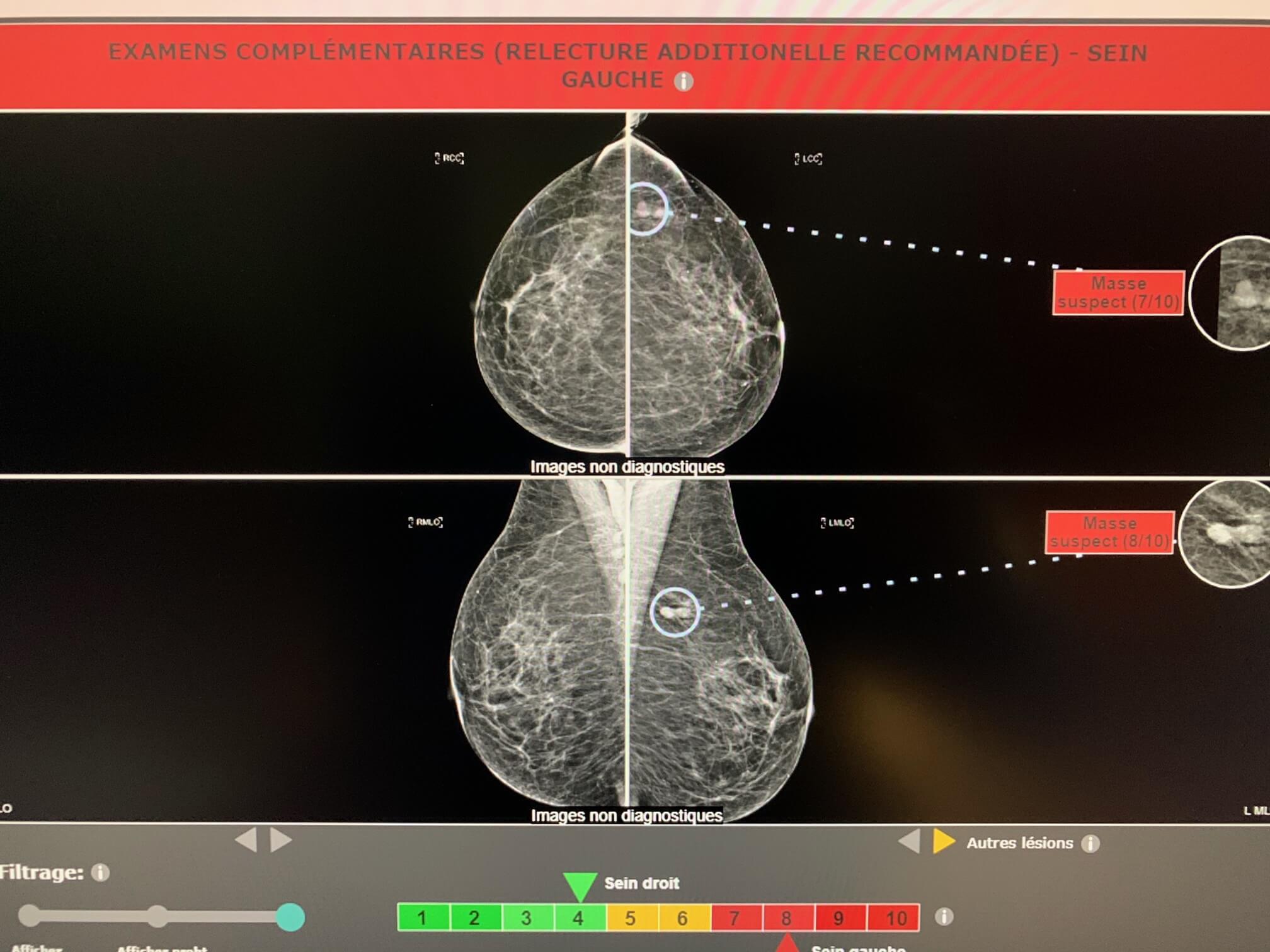
However, Mammoscreen™ points out these lesions and puts a red flag.
The mammogram is nevertheless interpreted as benign and sent for a second reading (in France, negative screening mammograms are verified by a second reader).
The second reader asks for an additional ultrasound and a spot compression in front of the slight increase in lesions between 2018 and 2020.
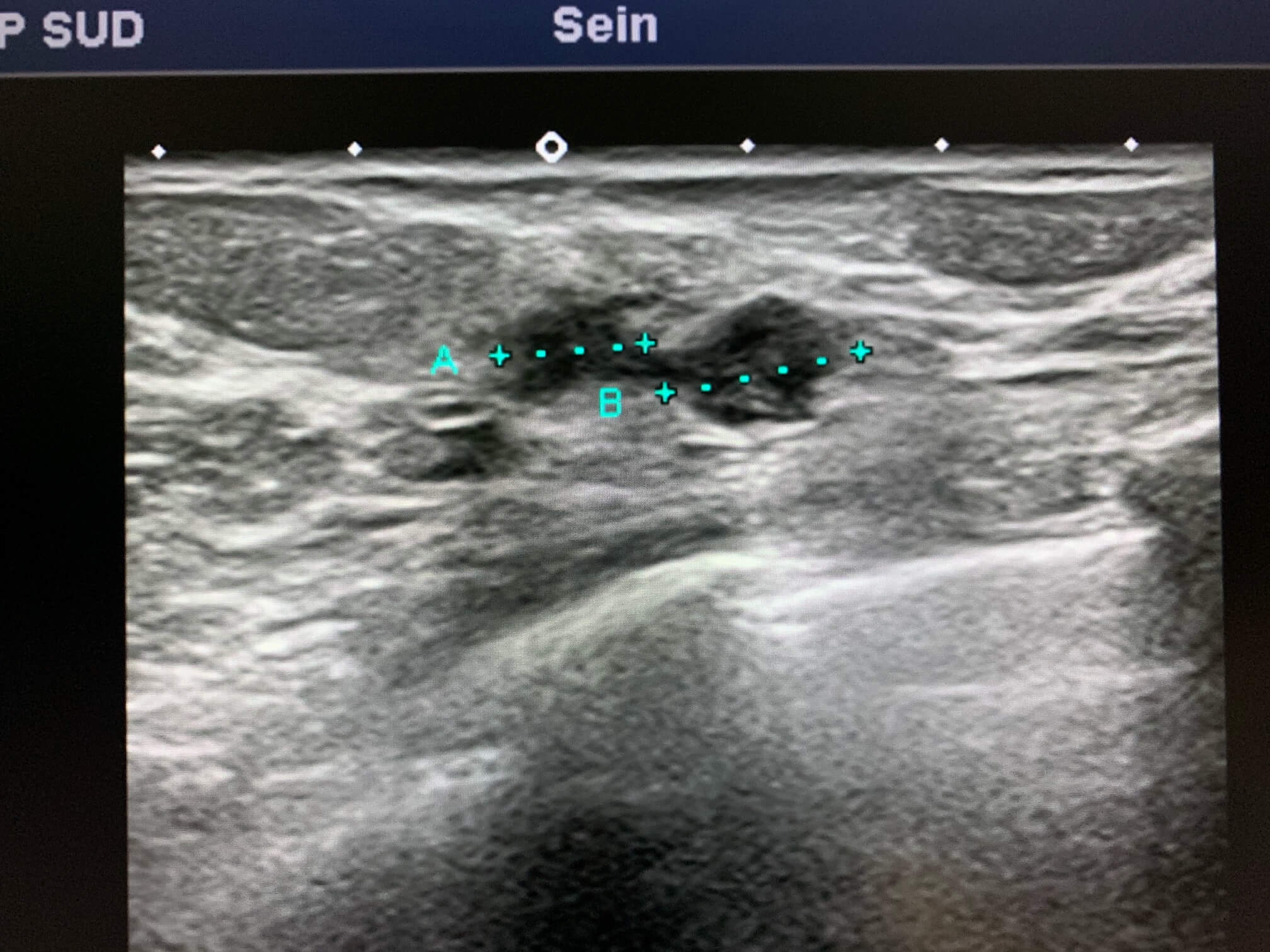
The ultrasound finds 2 rounded nodular formations next to each other.
A biopsy is performed and reveals an intra ductal carcinomatous proliferation and some fragments with infiltrating carcinomatous structures.
Conclusion:
- Be careful of pseudo-stability
- For countries without a second reading, MammoScreen could definitely help to prevent false negative interpretation.

*Case from the practice of Dr. Le Van An.