Case of the Week (week 35, 2022)
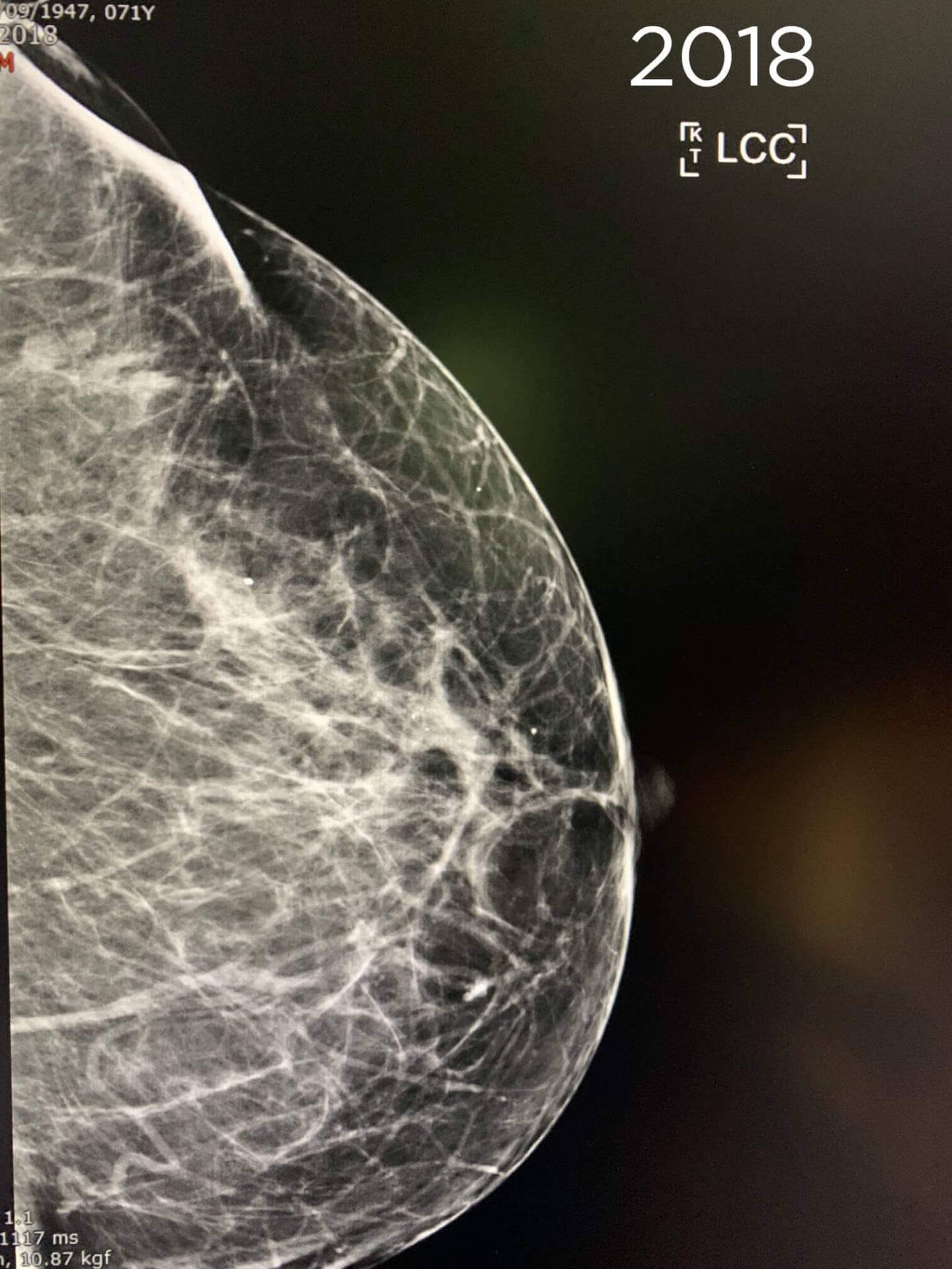
Screening mammography. 73-year-old patient
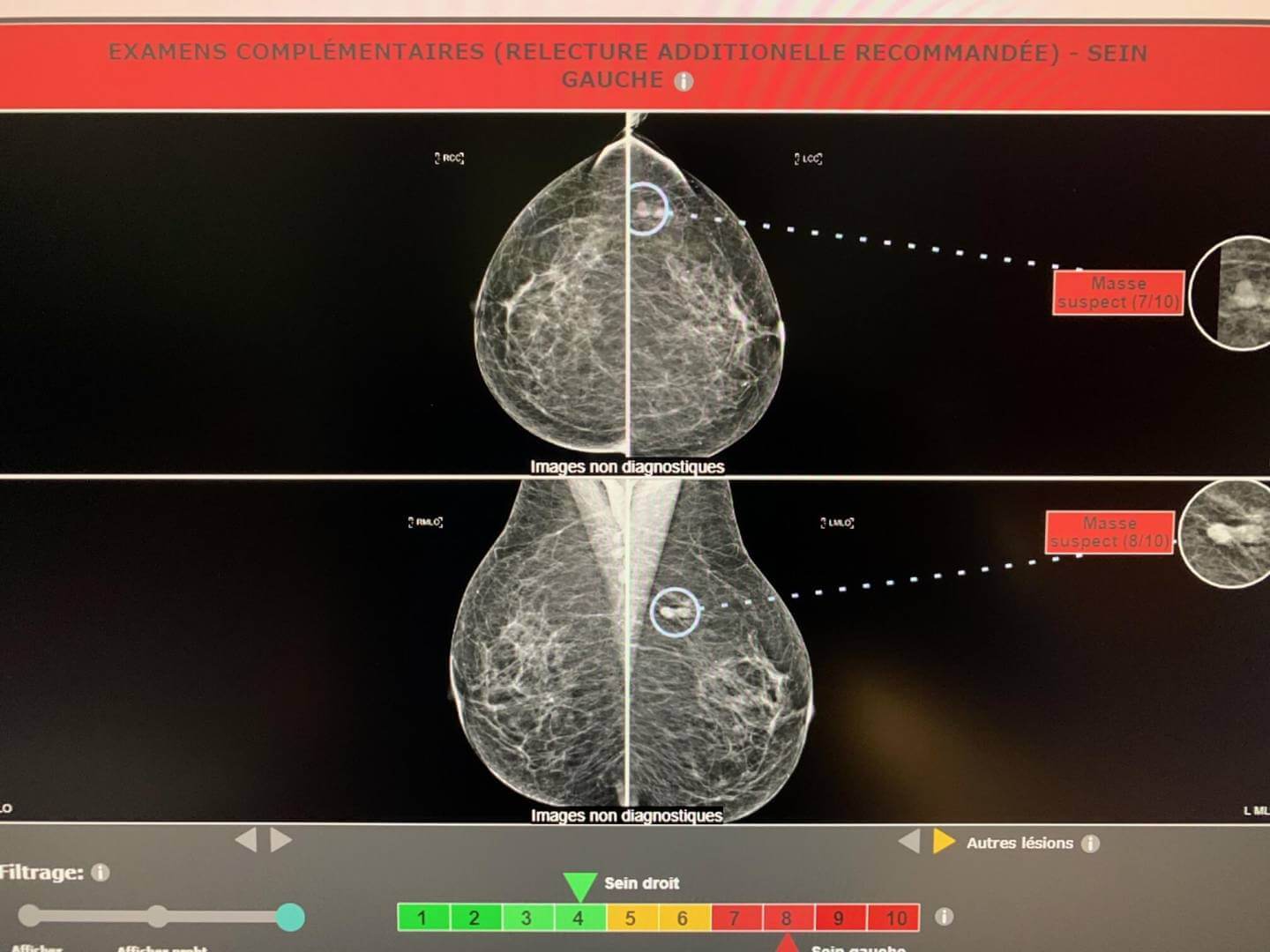
The exam is compared to the 2018 exam, there are 2 adjacent opacities in the left axillary extension present on the 2018 exam evoking intramammary lymph nodes.
Yet Mammoscreen® points to these lesions and puts on a red flag.
Mammography is still considered benign and sent for second reading.
The second reader asks for an ultrasound supplement and a localized image before the increase in lesions between 2018 and 2020.
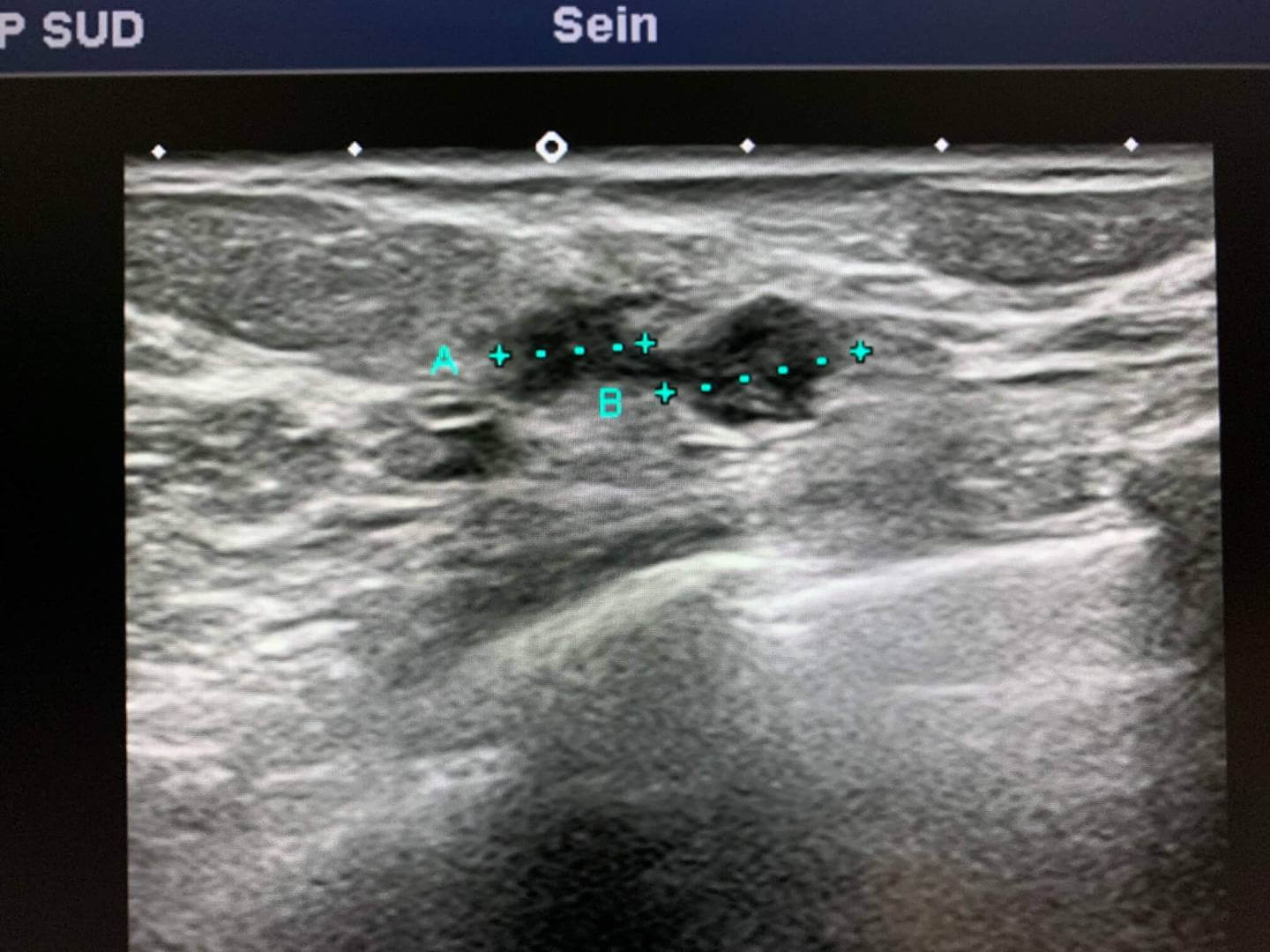
The ultrasound finds 2 rounded nodular formations next to each other.
A biopsy is performed and shows an in situ carcinomatous proliferation and some fragments with infiltrating carcinomatous structures.


*Case from Dr. Le Van An