Case of the week (week 14, 2022)
BILATERAL SCREENING 3-D MAMMOGRAM
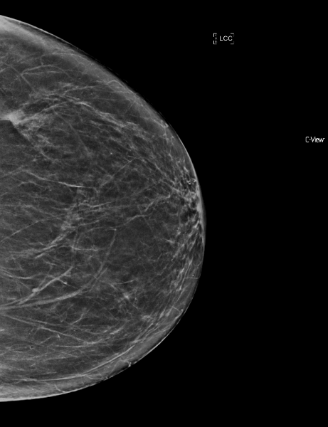
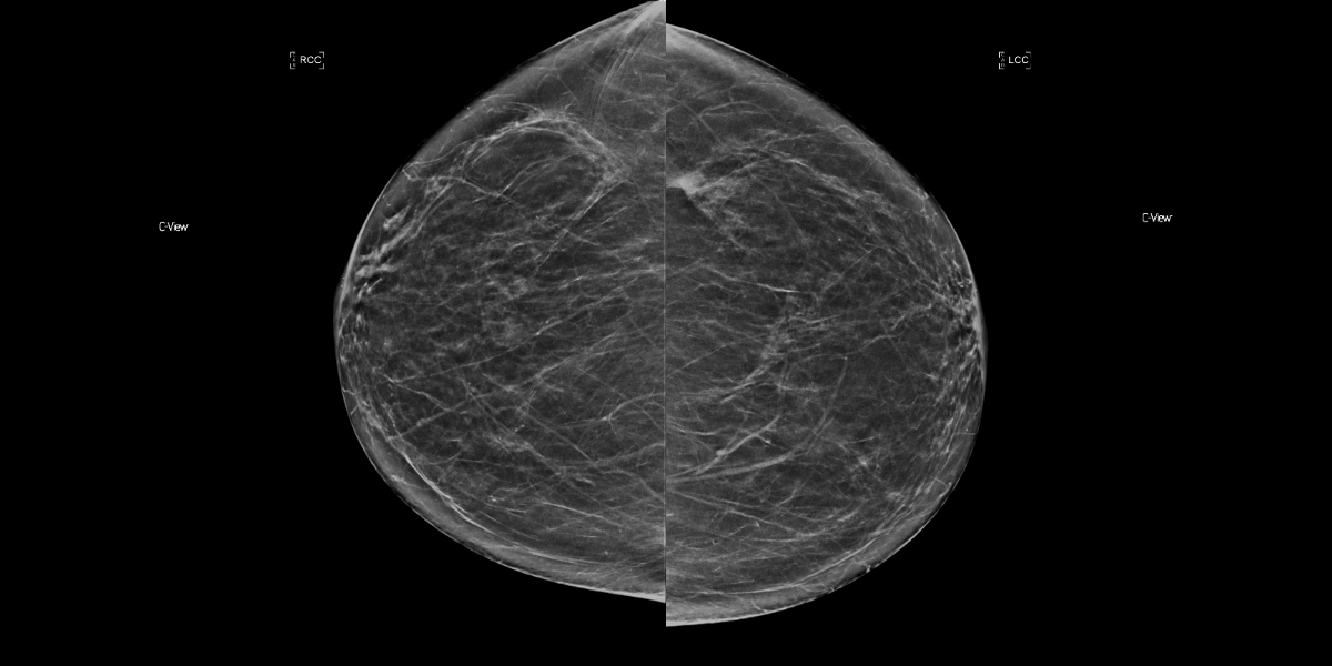
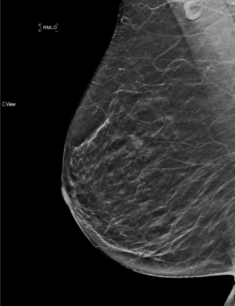
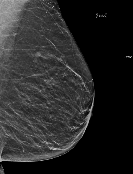
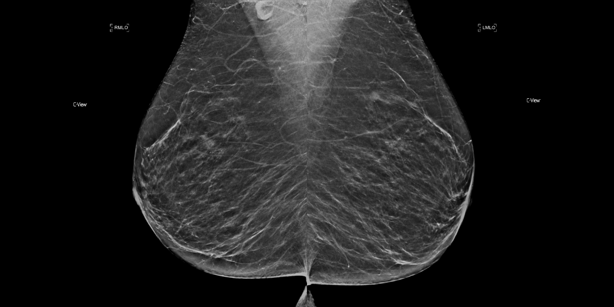
The breasts are almost entirely fatty. There is a lobular asymmetry within the upper outer aspect of the left breast posteriorly now present. Remainder of the left and right breast is negative for suspicious findings.
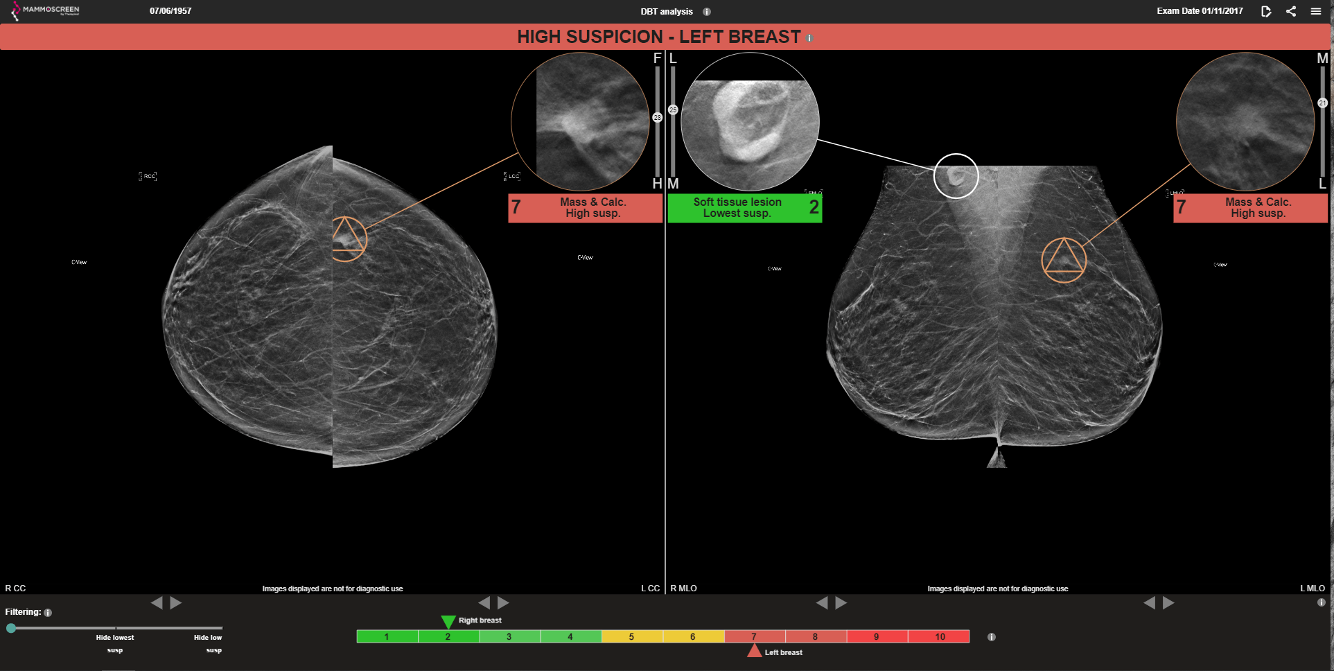
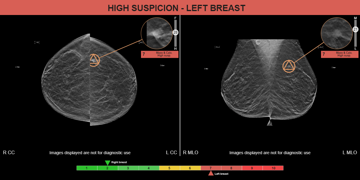
MammoScreen®: A collocated mass with micro calcifications is detected in both the LEFT CC and LEFT MLO. The system gave it a MammoScreen Score™ of 7. Detection of the lymph node observed in the RIGHT MLO was accurately categorized with a score of 2 – lowest suspicion.
MAMMOGRAM FINDINGS: There is a new small mass in the upper outer left breast measuring approximately 6 mm with irregular margins corresponding to the focal asymmetry noted on the screening mammogram. There is no associated distortion. There are a few scattered micro calcifications in the upper outer left breast which remain unchanged.
ULTRASOUND FINDINGS: Ultrasound performed of the upper outer left breast. At the 2:00 position 7 cm from the nipple there is a small irregular hypo echoic round mass measuring up to 6 x 5 x 5 mm with internal Doppler flow. This is subtle, firm and mobile upon physical exam. Ultrasound of the left axilla demonstrates a single mildly prominent lymph node with a fatty hilum and central cortical thickening up to 5 mm.