Case of the week (week 6, 2022)
BILATERAL SCREENING 3-D MAMMOGRAM.
Positive family history postmenopausal cancer in an aunt. Past surgical history pertinent for excisional biopsy right upper outer quadrant in 2002 which was benign.
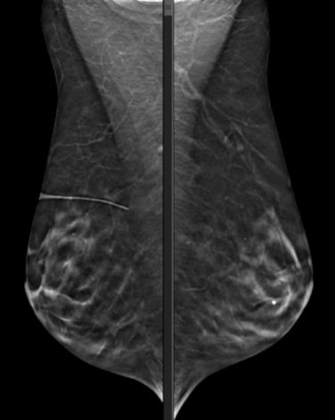
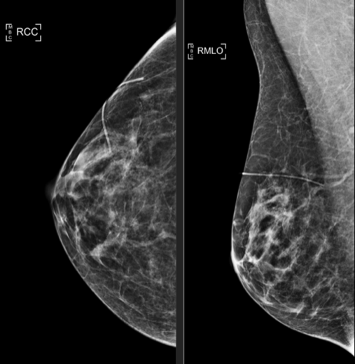
There are stable underlying dystrophic calcifications. In the upper inner RIGHT breast, there are new clustered pleomorphic, linear, and punctate microcalcifications in the middle depth, with no definite associated mass. These calcifications are not seen on prior mammograms.
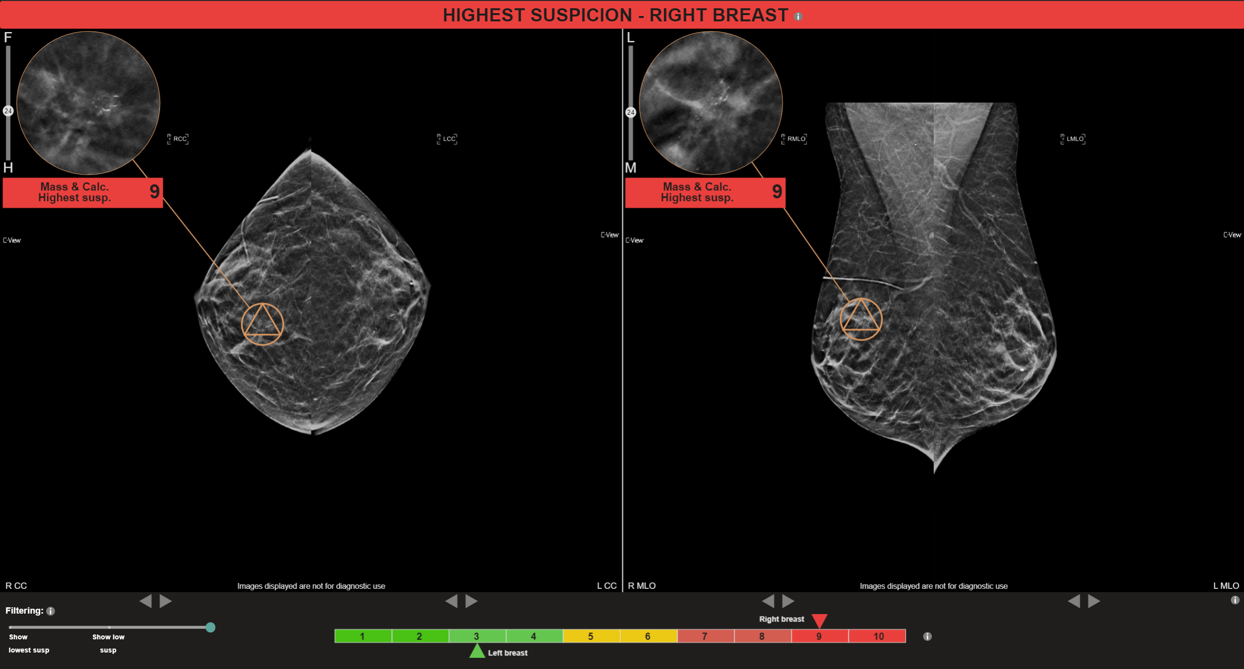
MammoScreen®: Co-located soft tissue lesion with calcifications identified in the RIGHT CC and RIGHT MLO views with a MammoScreen® Score of 9 supporting observation.
Recommendations: Unilateral RIGHT digital recall diagnostic mammogram.
Follow up diagnostic mammogram opinion: There is scattered fibroglandular tissue in both breasts. No new suspicious masses. There are some new calcifications in the right upper inner quadrant. These cover an area of approximately 3 cm x 2 cm. Many of the calcifications are irregular. Several branching calcifications. Calcifications have a somewhat ductal distribution. No associated mass or distortion. Findings are concerning for intermediate grade DCIS. Nodular asymmetry and adjacent punctate calcifications lateral right breast are stable since 2012 and are benign.