Case of the week (week 7, 2023)
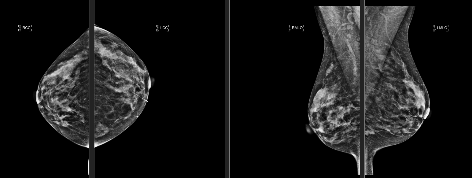
BILATERAL SCREENING 3-D MAMMOGRAM.
The breasts are heterogeneously dense. There is an irregular group of calcifications medial right breast. There is an area of potential architectural distortion immediately posterior medial to the calcifications on the right as well. In the left breast there is a well-circumscribed nodular density laterally, possible cyst. There is a micro lobulated nodular density far medial left breast
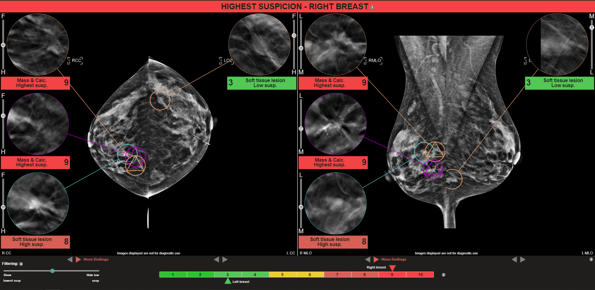
MammoScreen®: Two co-located masses with calcifications identified in the RIGHT CC and RIGHT MLO views with a MammoScreen Score™ of 9. One co-located soft tissue lesion in the RIGHT CC and RIGHT MLO with a Score of 8 all of which support observations. In addition MammoScreen identified the benign looking cyst in the left breast and gave it a low score of 3.
Recommendations: Bilateral diagnostic mammography with ultrasound.
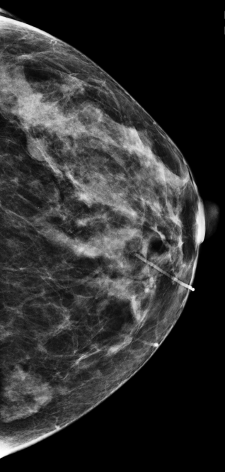
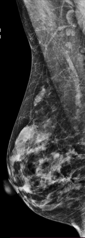
MAMMOGRAM FINDINGS: There is a well-circumscribed 10 mm mass in the outer left breast, likely a cyst. There is asymmetric breast parenchyma inferiorly and medially, previously evaluated and demonstrated by normal breast tissue. There are numerous narrowing sized slightly irregular rounded calcifications in the medial right breast. Calcifications are located predominantly anteriorly; there is posterior and lateral extension of the calcifications over a length of 4 cm. There is also associated architectural distortion. There may be a mass present.
ULTRASOUND FINDINGS: The inner right breast at the 9:00 position, 8 cm from the nipple there is a 1.7 x 1 0.6 to 0.5 cm elongated cystic mass or duct but no solid mass. At the 2:30 position, 5 cm from the nipple there is a 1.0 x 0.6 x 1.4 cm irregular, shadowing hypoechoic solid mass with internal vascularity. At the 3:00 position, 4 to 5 cm from the nipple there is a 2.5 x 1.7 x 0.8 cm irregular marginal shadowing hypoechoic mass containing calcifications and internal vascularity. The masses are 2.3 cm from each other.