Case of the week (week 5, 2022)
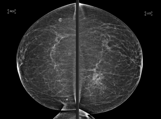
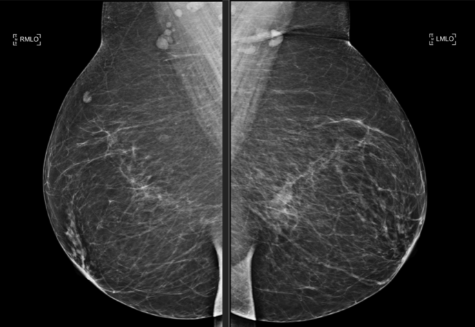
BILATERAL SCREENING 3-D MAMMOGRAM. symmetric densities observed in the posterior medial LEFT breast parenchymal margin. No mass, suspicious calcifications, architectural distortion or skin change in either breast observed.
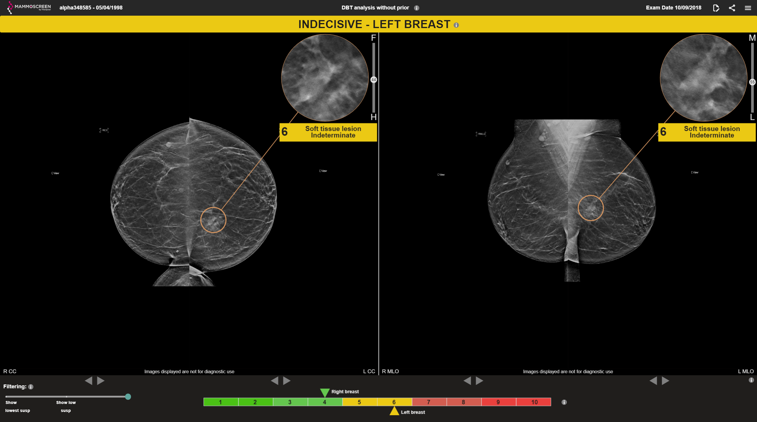
MammoScreen®: Co-located soft tissue lesion identified in the LEFT CC and LEFT MLO views with a MammoScreen® Score of 6 supporting observation.
Opinion: LEFT breast appears to have asymmetric density of an uncertain significance. Recommendation to perform a LEFT breast ultrasound to confirm.
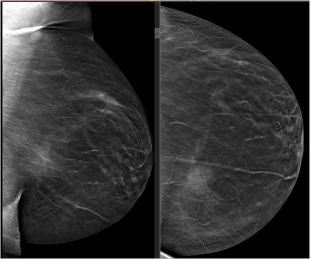
Ultrasound: Within the LEFT breast, 8 cm from the nipple at the 10:00 position, a lobular hypoechoic mass was identified measuring 1.2 x 1.6 x 0.7 cm in size. It has a parallel orientation. This correlates with the posterior mass observed on both the mammogram as well as the MammoScreen detection in the LEFT breast.