Case of the week (week 47, 2020)
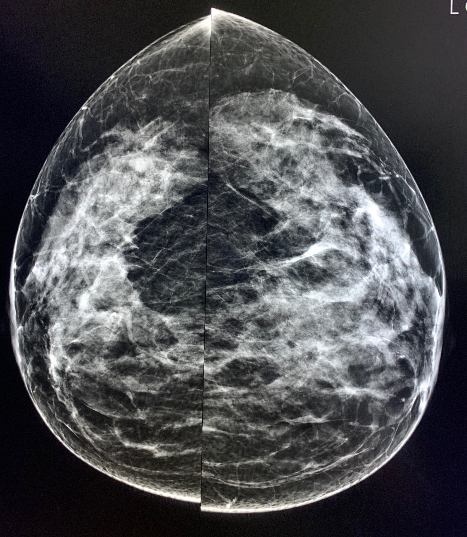
This 50-year-old patient had her first mammogram as part of a screening campaign.
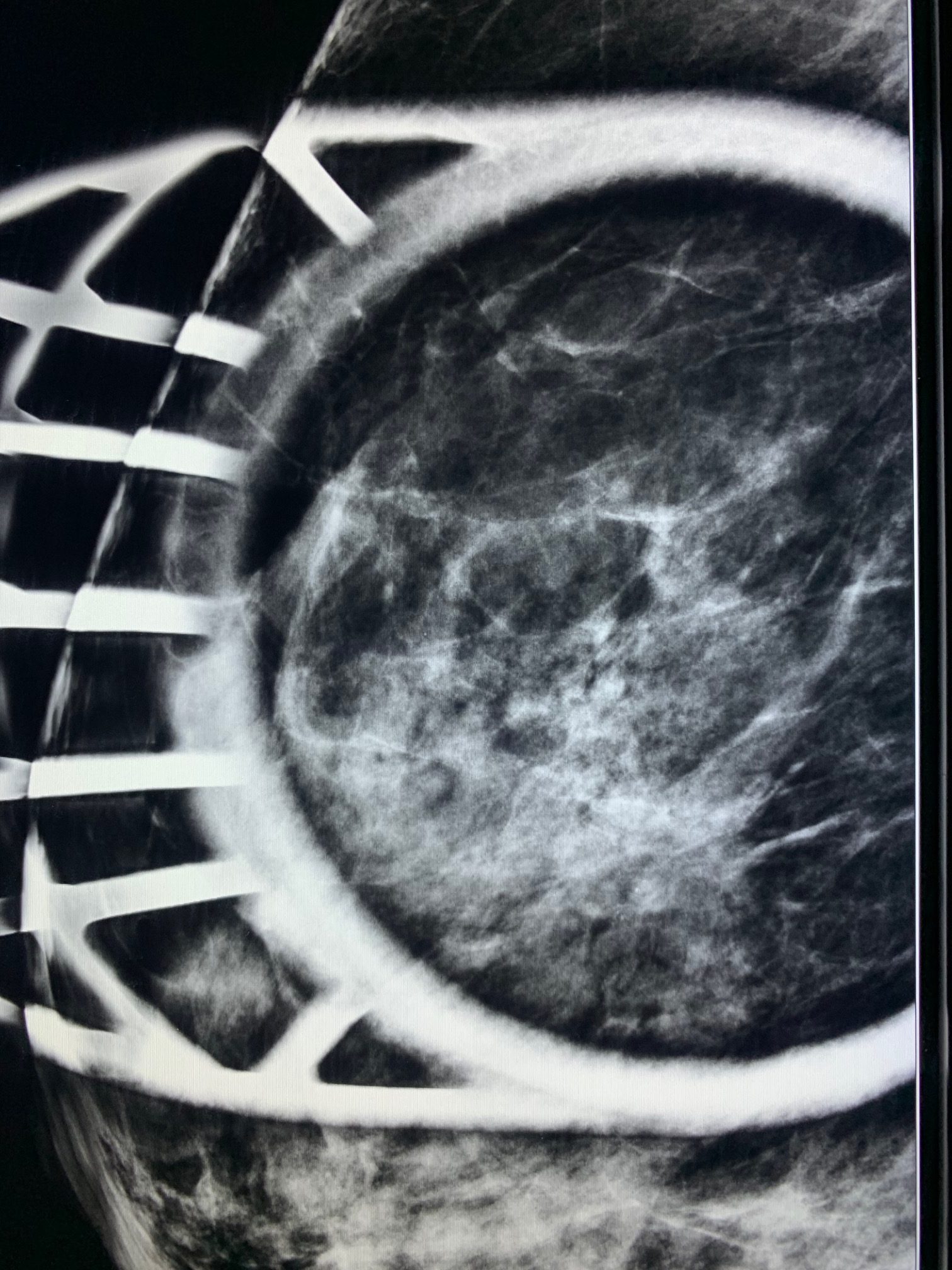
At first glance, the mammogram seemed normal. Tomosynthesis performed from the front showed a possible distortion, but distinguishing between a crossing of fibers and a pathological image was not easy (In hindsight it is more obvious.
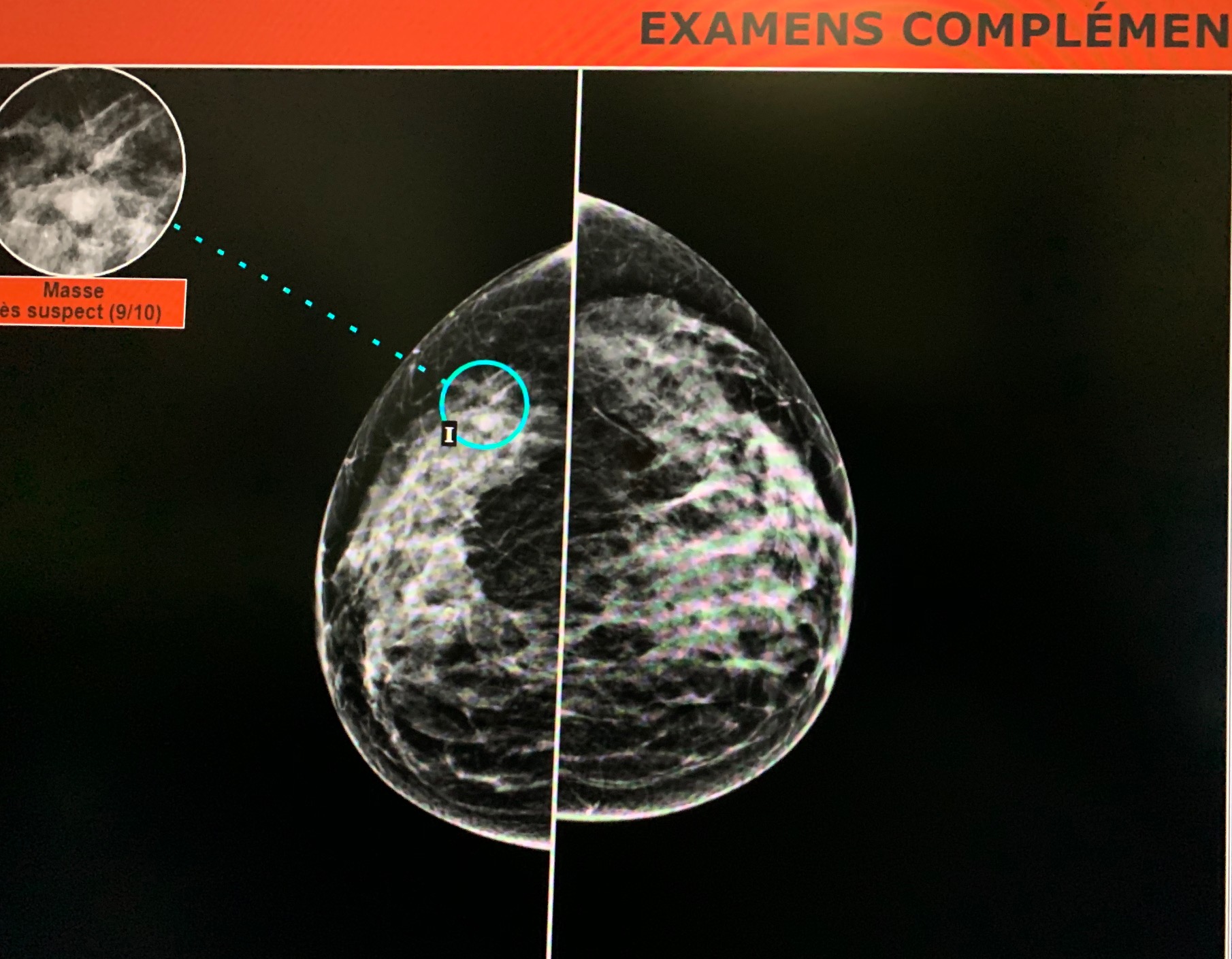
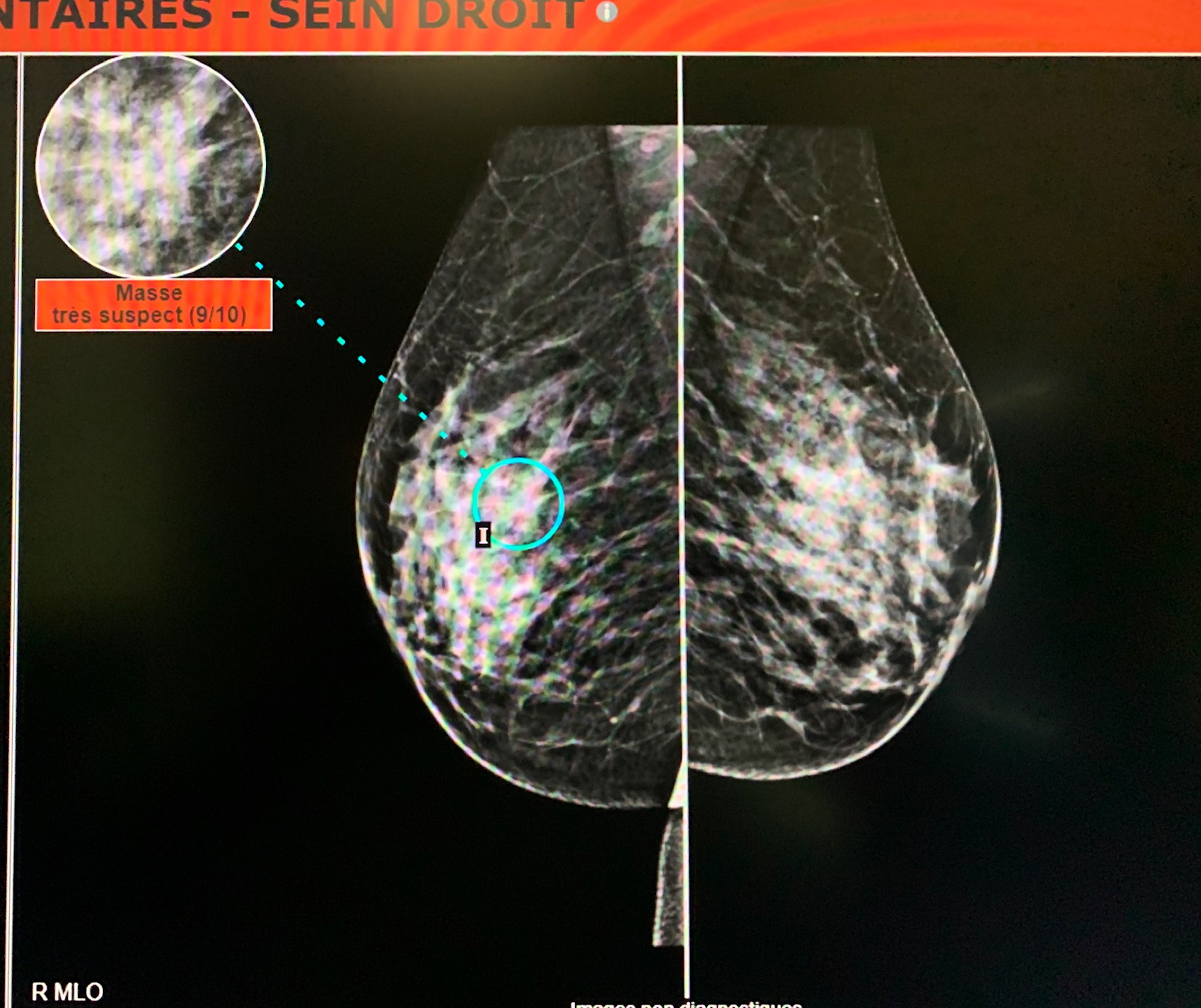
When I analyzed the case with an AI-based software MammoScreen™, I saw that the software pointed out an external retromammelonal zone.
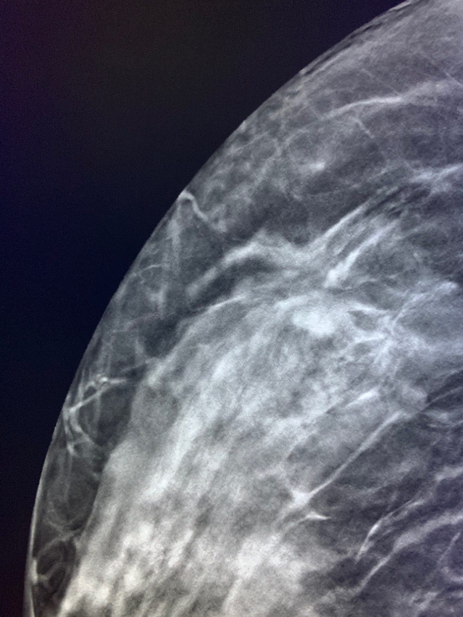
I made the front and profile spot compression views, but it did not bring out a spiculated image.
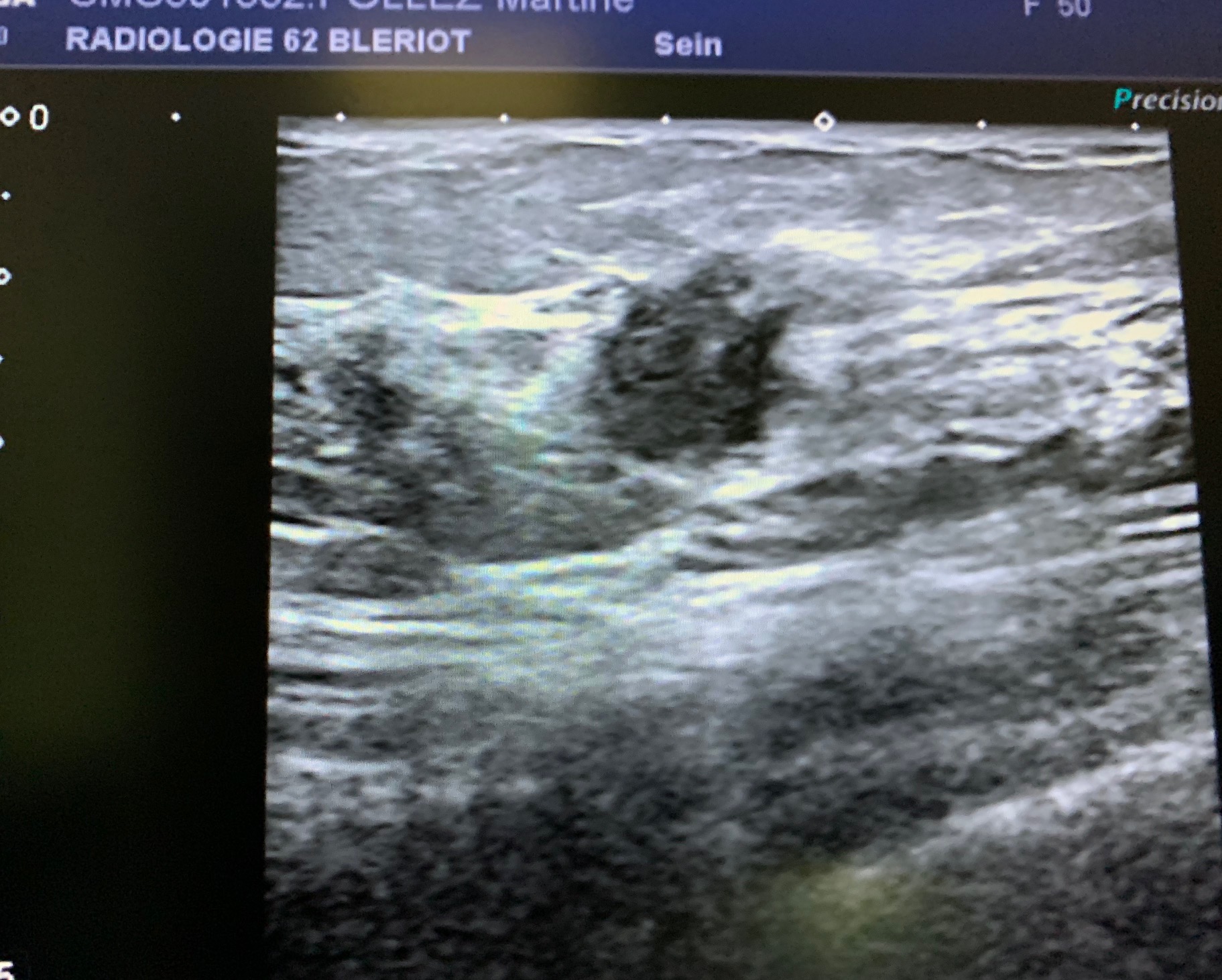
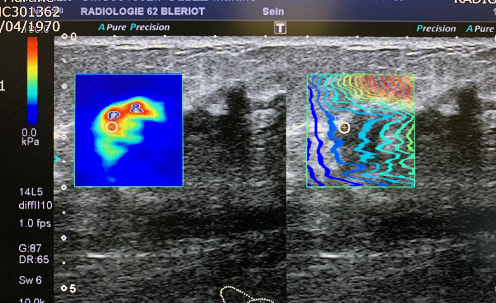
As MammoScreen™ highlighted the case in red, I thought that as a precaution, I should continue. Therefore, I made an ultrasound scan that highlighted a hypoechogenic image with irregular contours, with zones of rigidity under elastographic study, located at the union of the external quadrants of the left breast.
Ultrasound shows a hypoechoic mass with irregular contours and elastographic rigidity.
Breast biopsy: infiltrating carcinoma of luminal HER2+ type.

*Case from the practice of Dr. Le Van An.