Case of the Week (week 43, 2021)
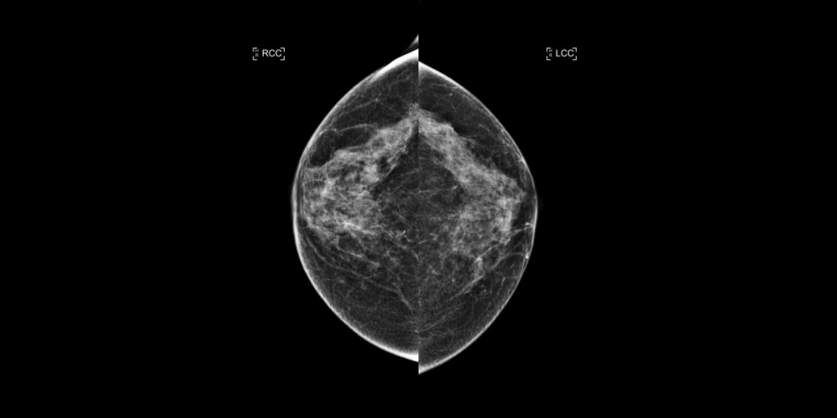
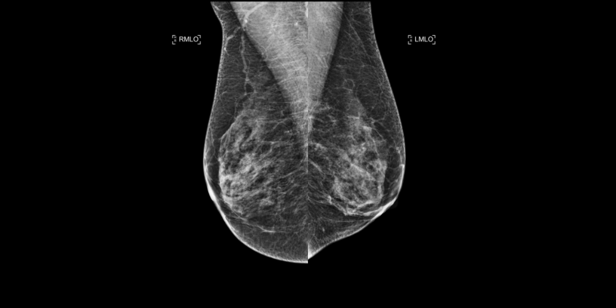
53-year-old female, test mammogram.
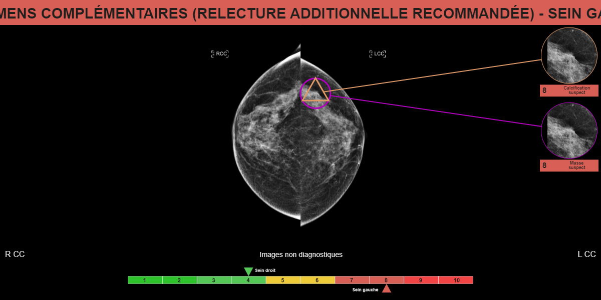
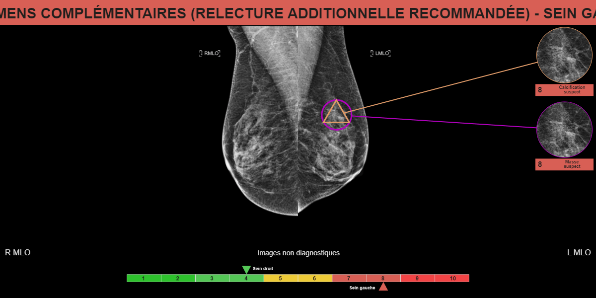
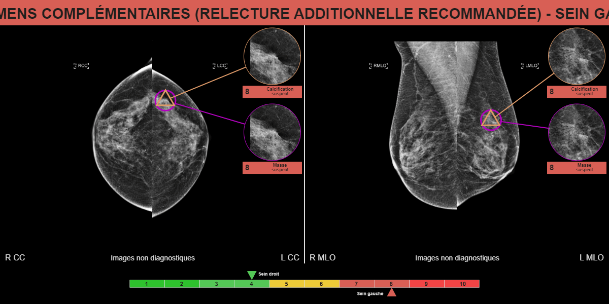
Mammoscreen points to increased opacity of the left breast QSE.
The localized image shows an opacity with irregular and spiculated contours.
Breast ultrasound confirms 7 mm irregular hypoechogene formation of the left breast QSE.
The microbiopsia finds an atypical epitheliosis.
Faced with the discrepancy between imaging and histology, a macrobiopsy is performed and confirms an infiltrating tubular carcinime and lesions of ductal carcinoma in situ.