Case of the Week (week 3, 2025)
During a screening mammogram, the radiologist identified a patient with a small 6 mm cancer located in the external part of the right breast. This initial discovery led to a recommendation for a biopsy in order to conduct further assessment to determine the nature of this abnormality.
Before biopsy, a complementary tomosynthesis assessment was performed using MammoScreen in parallel. Tomosynthesis revealed an additional lesion in the lower right breast area, which was confirmed by ultrasound.
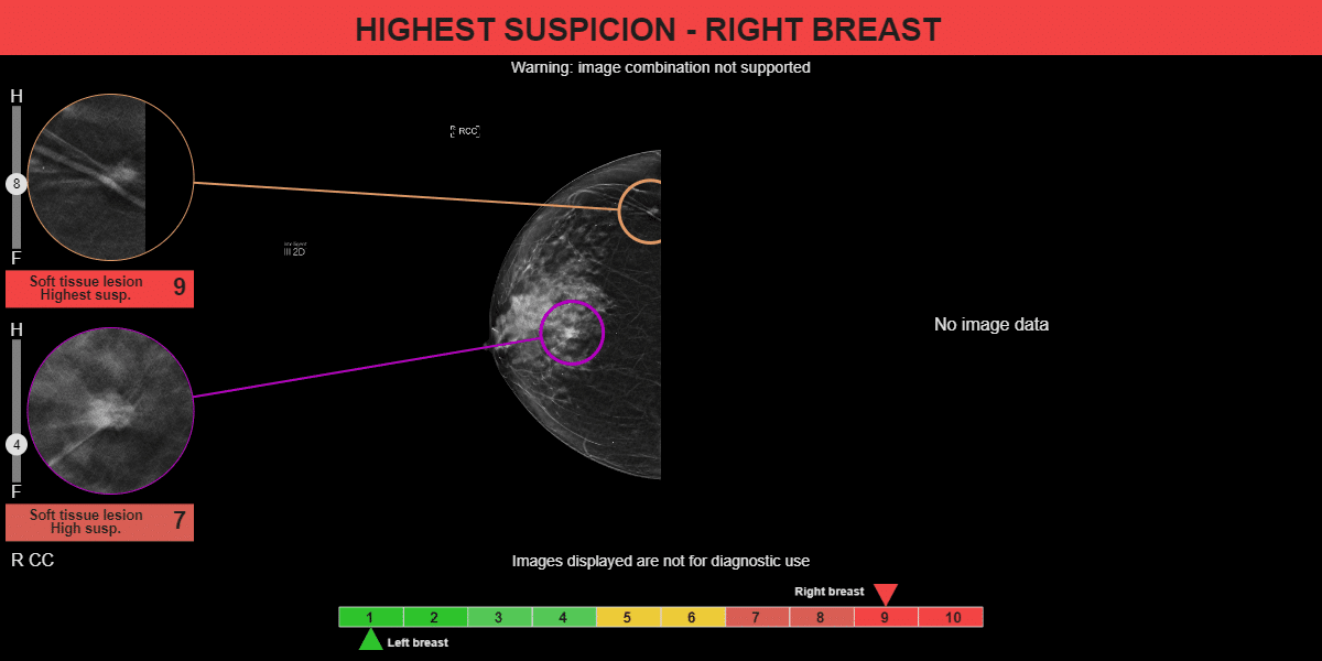
The results of MammoScreen show two distinct lesions on the patient’s right breast.
The first lesion detected is a very suspicious mass, which was given a score of 9 by MammoScreen. The second lesion identified by MammoScreen is a mass with a score of 7.
The MammoScreen analysis confirms the presence of both suspicious lesions and would have allowed the first reader to not miss the additional lesion.